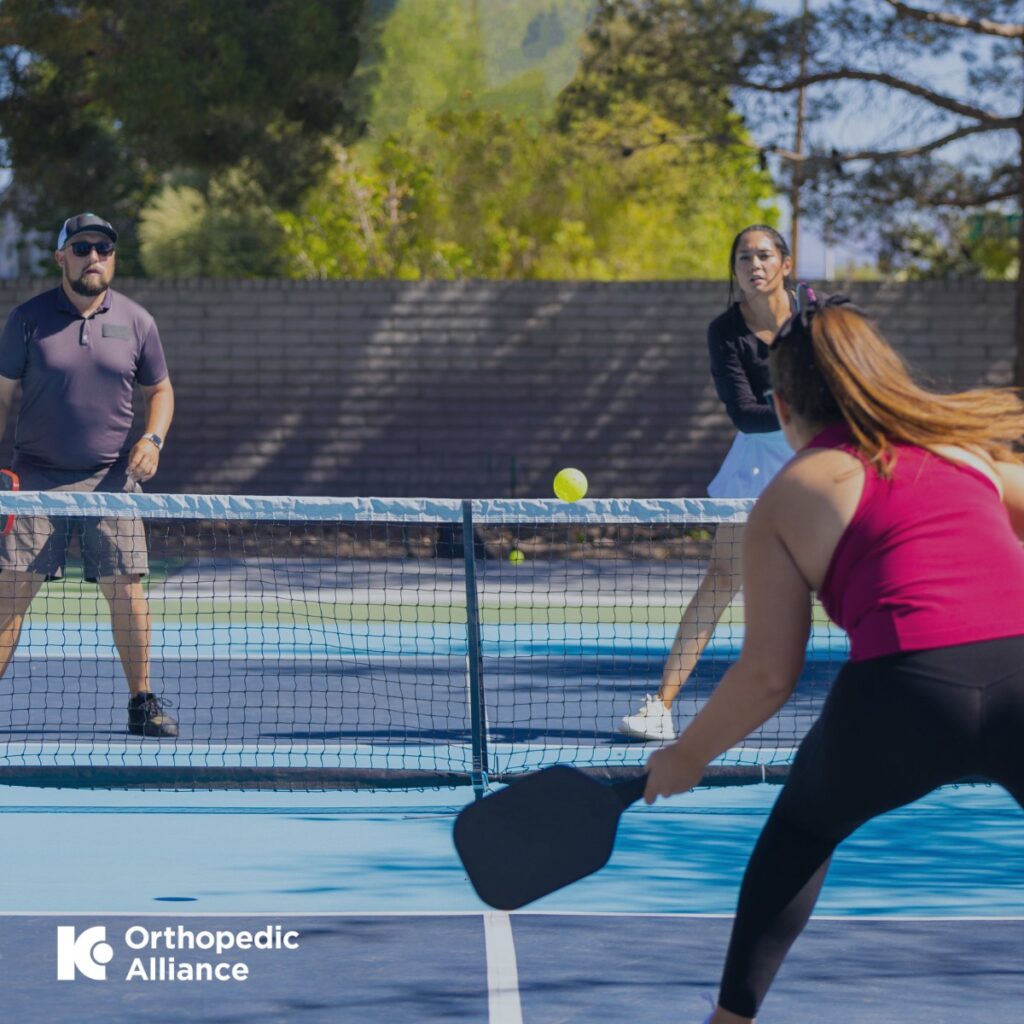
Nerve injuries require unique methods to fully assess the presence, extent, and location of abnormality. Your physician will individualize and guide management plans based on these factors, thus management for one person can vary from management of another person.
Dr. Moore, our newest PM&R physician at APEX Orthopedics & Sports Medicine, is both specifically trained in electrodiagnostic studies and ultrasound to evaluate the nerves and determine the most beneficial nerve injury treatment for patients.
Read on to learn more about various nerve injury evaluations as their potential foundation for future pain management.
Common types of peripheral nerve injuries
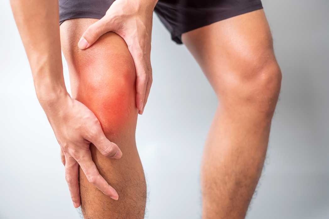
“Nerve injury” is a broad term that encompasses a variety of conditions. Therefore, before coming up with a treatment, it’s important to understand which type of nerve injury you have.
The nervous system can be thought up as a series of electrical wires and connections from the brain to the spinal cord (also known as the central nervous system) and from the spinal cord to your body (known as the peripheral nervous system).
The peripheral nervous system starts as large bundles of nerves that exit the spine (called spinal nerves). Most nerves then undergo a series of reorganization, splitting and combining in specific patterns at locations called a nerve plexus, and eventually separate into individual peripheral nerves (like the ulnar nerve and common peroneal nerve). Each peripheral nerve has specific functions that may include serving (or innervating) certain sensory regions of the body and certain muscles. Abnormalities, diseases, or injury can occur at any level of this nervous system organization. In general Dr. Moore sees and manages two common patterns of nerve injury.
- Nerve entrapments
- Radiculopathies

Nerve Entrapments
Nerve entrapments are a type of mononeuropathy, which means a focal injury to a single peripheral nerve at a specific area in an extremity. Entrapment neuropathies (also called compression neuropathies) include diagnoses like carpal tunnel syndrome and cubital tunnel syndrome. In an ideal anatomical course, nerves travel in smooth channels or cushioned spaces between muscles, ligaments, and bones. When those spaces around a nerve narrow, it can cause nerve compression and irritation, leading to pain and discomfort. Nerve compression can be due to various factors such as:
- Specific anatomy: your body may naturally have narrower tunnels for the nerve to travel through or normal anatomical variants (accessory muscles, unique neuroanatomy) that may predispose a nerve to be compressed at certain areas.
- Trauma: Acute trauma can cause nerve damage, such as a displaced bone fracture pressing on a nerve.
- Repetitive activities: Repetitive microtrauma can cause nerve irritation as well, particularly in locations where a nerve is superficial and prone to compression from external forces.
- Injuries: Certain injuries can cause scarring of your soft tissues that can tether or entrap a nerve passing nearby.
- Osteoarthritis: Bony enlargement from arthritis occasionally can cause nerve entrapments.
Nerve compression leads to reduced blood supply (ischemia), and without proper blood supply, the nerves cannot work properly to send and receive messages. The specific symptoms of nerve compression depend on the specific nerve involved and what it innervates. Common symptoms of nerve compression include:
- Pain
- “Pin-and-needles” sensation
- Numbness
- Muscle weakness
Radiculopathy
Radiculopathy is the medical term for irritation of a spinal nerve (the large bundles of nerves that exit the spine). Cervical radiculopathy is when irritation occurs to a cervical spinal nerve at the neck level, while lumbosacral radiculopathy (commonly referred to as “sciatica”) is irritation to a lumbosacral nerve at the low back level. They are commonly referred to as “pinched nerves,” though that terminology has a much more aggressive connotation than what generally is occurring at the anatomical level.
Radiculopathies typically occur from disc bulges/herniations or degenerative changes in the spine causing narrowing and inflammation around where the spinal nerve exits. Less commonly radiculopathy may be caused by spine tumors or infectious causes.
There are a lot of important complexities in the history and exam in someone with radiculopathy that affects management, so it is always important to visit a doctor if you think you may have symptoms concerning radiculopathy. Classically, radiculopathy causes radiating extremity pain (pain radiating down the arm in cervical radiculopathies, pain radiating down the leg in lumbosacral radiculopathies) more so than frank neck or back pain. This is because often with nerve pain, one’s body interprets the nerve irritation as coming from where the nerve innervates in the extremity rather than focally where the nerve irritation is actually occurring. The pain may worsen with certain activities. Other potential symptoms of radiculopathy include numbness, tingling, or focal weakness in the affected limb.

Nerve injury evaluations and methodologies
Two common, and often complimentary, ways to evaluate nerves are with electrodiagnostic studies and ultrasound.
Electrodiagnostic studies
Electrodiagnostic studies help to measure the function of the nerves and consists of two portions:
- Nerve conduction study (NCS): sending small shocks down a nerve to assess its ability to carry information
- Electromyogram (EMG): placing small needles in the muscles to evaluate messages from the nerves for abnormalities
The information received from electrodiagnostic studies is similar to piecing a puzzle together to solve a problem. Therefore, the length of the study depends on two factors:
-
- The complexity of the question: For example, is your study simply meant to rule out carpal tunnel syndrome or is it also to evaluate for underlying radiculopathy or multiple other peripheral nerve entrapments in the entire upper limb? The latter will take longer to perform than the first.
- How obvious or complex the findings are: In some cases, your physician may require more information from other nerves to reveal a specific pattern that suggests a specific diagnosis.
With those factors in mind, an average electrodiagnostic study takes about 30 to 90 minutes to complete.
The NCS shows whether or not electricity is properly conducted through a specific area of the nerve. You can think of this as checking an electrical wire for damage. As small shocks hit a portion of the nerve, the response is measured upstream and downstream to gauge factors such as velocity and amplitude. From there, your physician can compare those values to “normal values” for appropriateness.
The EMG portion of the study uses acupuncture-sized needles into the muscle to listen to the electrical response of the nerve to that muscle. When Dr. Moore performs an EMG, she first listens to the nerve at rest (when it should not be firing) then she listens when you flex (or activate) a muscle. The idea is similar to ensuring the electrical wiring connected to a light switch works properly to turn on a light.
Lastly, Dr. Moore interprets the NCS with the EMG along with the clinical history and examinations to come up with the impression of the study (e.g. confirming carpal tunnel, or ruling out radiculopathy).
Electrodiagnostic studies are unique studies, and often patients have less familiarity with what to expect with them then compared to more common evaluations like X-Rays and blood draws. Patients generally tolerate both portions well, though they may cause some discomfort at times. However, Dr. Moore will be right beside you, and you may take breaks as needed. If you cannot tolerate the exam, she will stop.
Ultrasound
Ultrasound is an imaging modality that can be used to evaluate the structure of the nerve and potentially localize sites of compression, entrapment, or other injury for targeted management. Understanding the nerve injury and the extent of the injury leads to discovering which treatment options will best suit the patient. Of course, each treatment is fully individualized to each person and his or her goals.
Dr. Moore may use diagnostic ultrasound in conjunction with electrodiagnostic studies when needed to supplement the study. Keep in mind that the electrodiagnostic highlights the function of the nerve, while the diagnostic ultrasound shows the structure of the nerve.
Here is how the ultrasound works to reveal a nerve’s structure:
Ultrasound is a noninvasive imaging modality that uses sound waves instead of ionizing radiation to visualize anatomical structures in real time. The ultrasound transducer (or probe) is a handheld device that emits high-frequency sounds waves through the soft tissues in front of it. The sounds waves reflect back to the probe in varying degrees, and those reflections (or echoes) are then reformatted into an image in real time showing the structures you are looking at. This process is very similar to how boats and submarines use sonar technology for travel.
During a diagnostic ultrasound of a nerve, the nerve itself will be imaged, measurements of nerve size may be taken and compared to the other extremity, and dynamic maneuvers may be performed such as scanning the nerve while you move to look for areas of intermittent compression.
Nerve Injury Management
If you are referred to Dr. Moore by another physician for electrodiagnostic testing or diagnostic ultrasound alone, she will provide a full summary of the findings and impression to your referring physician for further discussion on management plans. If the studies were performed as part of a full evaluation from an earlier meeting with Dr. Moore, she will then discuss with you the management options going forward and what path best aligns with your specific case. She is trained in a wide variety of conservative management approaches such as:
- Ultrasound-guided injections
- Hydrodissection procedures
- Medications for short-term pain relief (i.e. steroids)
- Long-term pain relief options that optimize nerve healing (i.e. platelet-rich plasma)
- Minimally invasive procedures (i.e. carpal tunnel release with ultrasound guidance)
The right treatment option depends on the underlying type and extent of the nerve injury/pathology. Dr. Moore understands when to refer a patient to a surgeon, when to refer to a neurologist, and when there is something she can offer to give you the best results.
Get to know Dr. Moore at APEX Orthopedics & Sports Medicine
Dr. Moore is our newest addition to the APEX team and one of the only two sports-fellowship trained PM&R physicians in the Kansas City area. With subspecialties in sports medicine and musculoskeletal ultrasound, Dr. Moore has the credentials and tools to treat anything from acute injuries to chronic overuse.
Her time at Mayo Clinic has ingrained the importance of using an evidence-based approach while remaining on top of the curve on cutting-edge medical advancements. In her practice, she utilizes therapeutic and diagnostic ultrasound-guided injections, regenerative and orthobiologic treatments, and advanced,minimally invasive tendon procedures such as Tenex.
The core of her success lies in proper diagnosis. An appointment with Dr. Moore includes the highest quality technology to pinpoint a patient’s underlying musculoskeletal conditions. From there, she can better understand which treatment option would be the best solution.
For a thorough nerve injury evaluation with Dr. Moore, call us at 913-642-0200 or schedule an appointment today.